| Lab Section | Serology |
| Test Description | Serum neutralization test for the detection of antibodies to the equine arteritis virus |
| Species | Equine |
SUBMISSION DETAILS
| Sample Type | Serum (1 ml) |
| Container | Red top tube or other sterile container without additive |
| Special Submission Instructions |
|
| Turnaround time | 4-7 business days |

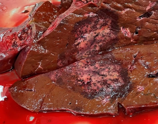
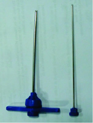 Using histopathology to definitively diagnose canine bone lesions is very important, since benign and neoplastic osteodestructive processes can be indistinguishable on radiographs and diagnosis greatly affects the treatment regimen. However, representative sampling of the lesion by bone biopsies (e.g., Jamshidi bone needle, Michele trephine) can be quite challenging. The center of the lesion is often necrotic, while the edges may simply be reactive changes of bone and periosteum. Sometimes, despite everyone’s best efforts, the tissue sampled is non-diagnostic. A retrospective study of VMDL cases demonstrates that neoplastic changes were not observed in 20 out of 61 (33 percent) primary bone tumor cases in dogs at the first bone-core biopsy attempt (Kuroki et al. Accuracy of bone-core biopsy for diagnosis of primary bone tumors in dogs. 2012 ACVP and ASVCP annual meeting. Paper #D-47). While it’s a good idea to prepare your clients for the possibility of an unsuccessful diagnosis, here are a few things you can do to improve your chances of getting a definitive diagnosis: